Canned Pickled Lettuce,Pickled Lettuce Canned,Pickled Lettuce Tin,Lettuce Canned ZHANGZHOU TAN CO. LTD. , https://www.zztancan.com
In osteoporotic animal studies, bilateral ovarian ablation in female rats is a more mature model that can successfully establish osteoporosis symptoms that mimic estrogen deficiency.
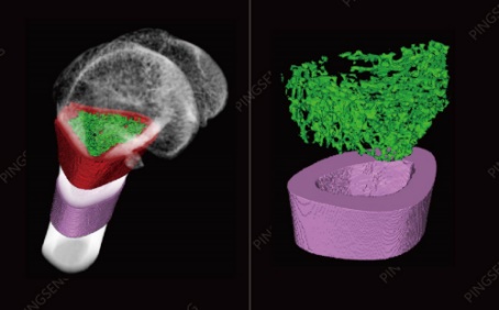
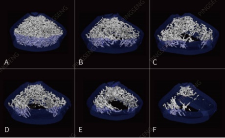
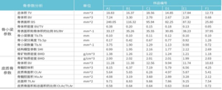
Example: 6 experimental rats with AF number were isolated femur samples, of which A was the normal control group, and B to F was the osteoporosis group (ovarian model) using different anti-osteoporosis drugs. The segmentation process is shown in Figure 1; the bone trabecular display in the region of interest is shown in Figure 2; the bone parameters of the ROI trabecular bone and ROI cortical bone are calculated as shown in Table 1. 
Osteoarthritis is characterized by subchondral bone sclerosis or cystic changes, bone hyperplasia at the joint edge, synovial hyperplasia, and narrowing joint space. 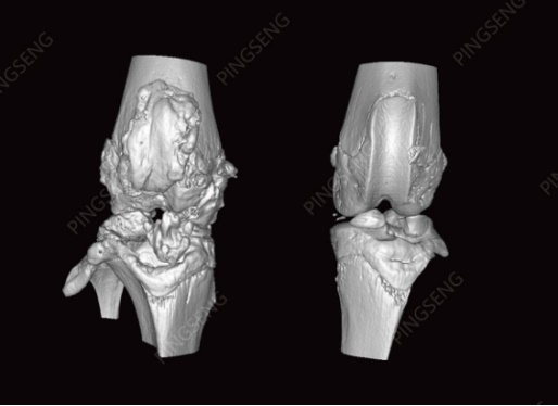

Experimental equipment: VENUS® Micro-CT
Chinese name: desktop high resolution micro CT
Model: VNC-100 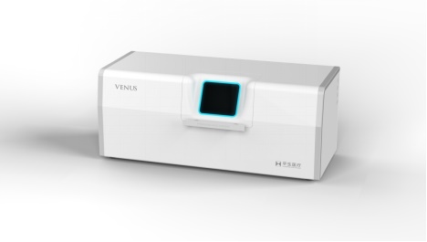
Application of micro-CT in bone imaging and quantitative analysis
Osteoporosis research
Micro-CT imaging is particularly important for the study of osteoporosis, particularly disease progression and therapeutic effects, as it is one of the few imaging techniques that provide information on bone mineral content and density. Measuring these changes through high-resolution micro-CT helps to develop therapeutic agents and understand the molecular mechanisms that control these processes.
Figure 1: Three-view and 3D location selection of the region of interest
Figure 2: 3D perspective display; segmentation extraction of trabecular bone and cortical bone in the region of interest
Figure 3: Differential display of trabecular bone performance in the normal group and different drug interventions in the AF group
Table 1: Calculation of bone parameters for ROI trabecular bone position (1.5 mm below growth plate, 2 mm long), ROI cortical bone position (5 mm below growth plate, 2 mm long)
Bone regeneration material research
In bone research on material implants, the usual goal is to examine osseointegration, the state of the bone around the implant. Micro-CT can provide 3D image data of the implant and surrounding bones and provide correlation analysis.
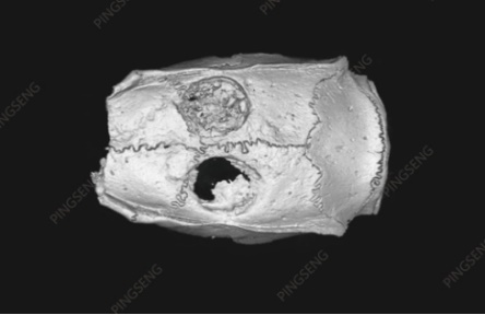
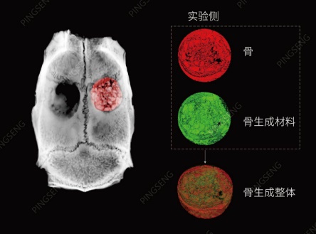
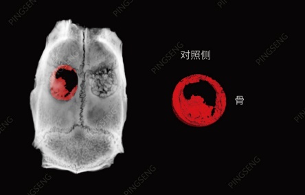
Figure 4: The repair effect of different treatments on the skull openings on both sides of the same rat. After opening the hole, the bone-forming material was added on one side (upper side of the figure), and the opposite side was used as the control side without any material (lower side of the figure); after a period of feeding and growth of the skull, the rats were examined by micro-CT. The skull scan was performed to observe the repair of the skull on both sides. It can be observed that the recovery of the skull on the side of the bone-forming material is better than that on the control side.
Figure 5: Segmentation of bone-forming materials and new bone by Avatar software
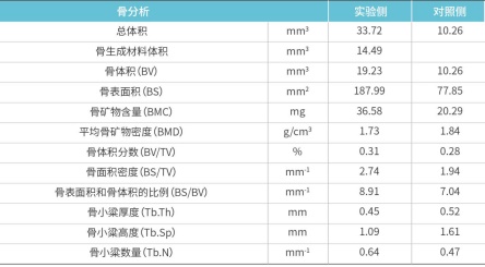
Table 2: New bone analysis on the material side and the control side
Osteoarthritis research
Establishing an animal model of osteoarthritis (OA) is an important way to find effective treatments for arthritic diseases. Micro-CT can detect small structural changes in the bone marrow and cortical bone, and has great advantages in evaluating small joints compared with other imaging methods. Micro-CT can be used to assess the small changes in cartilage bone in the progression of osteoarthritis, to assess bone mineral density and cartilage ossification to study the pathophysiology of osteoarthritis and the changes in calcareous deposits in cartilage.
Figure 6: Comparison of knee joints on both sides of the same rat. The upper side is modeled for osteoarthritis and the lower side is the normal control side. After a certain drug intervention, micro-CT was used to study the articular cartilage repair and arthritis progression, focusing on the articular surface and meniscus leveling and the volume change of the joint cavity.
Figure 7: As in this example, the knee joint cavity becomes larger due to bone hyperplasia at the edge of the joint. Divided and calculated by Avatar software, the figure (left) (red, arthritis), cavity volume 42.7mm^3; (right) (green, normal control) cavity volume: 23.3 mm^3.
Imaging software: Avatar 1.3 (Pingsheng Medical Technology)