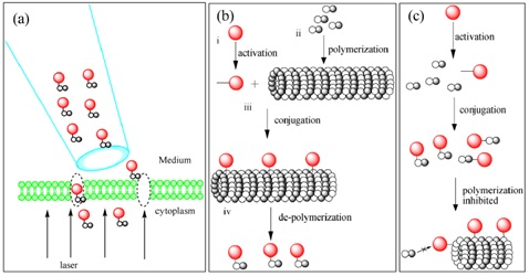
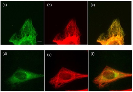
As a nano-fluorescent probe for inorganic synthesis, quantum dots have high fluorescence brightness and fluorescence stability, and are suitable for long-term observation and live tracing. Targeted delivery of quantum dots into the cytosol contributes to intracellular protein transient interaction studies, as well as long-term observations of dynamic cytological response mechanisms. At present, the methods for the delivery of quantum dots into cells are mainly divided into two categories: 1 assisted delivery strategy: the use of transmembrane peptides, polymer carriers, transfection reagents, etc. to achieve quantum dot delivery, but need to consider the quantum dots are endocytosed by cells. Release efficiency; 2 active delivery technology: including electroporation and microinjection, which will cause a certain degree of mechanical damage to the cells. The University of California's Shimon Weiss team used Photothermal nanoblade technology for intracellular delivery of quantum dot conjugates without regard to release efficiency after endocytosis, while avoiding mechanical damage. The researchers applied a laser pulse to the surface plasmon of the capillary tip to form nanosecond explosive bubbles in the immediate vicinity of the cell membrane. These bubbles instantaneously form slits or holes in the cell membrane; at the same time, pressure is applied to the capillary to form a strand. In the transient flow, the quantum dot conjugates then enter the cytosol (Fig. 1a). Compared with the traditional microinjection method, the contact between the photothermal nanosheet and the cell membrane is milder, avoiding mechanical damage to the cells. The above researchers applied this technique to the delivery of tubulin-quantum dot conjugates to achieve real-time observation of microtubule skeletons in living cells. As shown in Figure 1, the researchers improved the coupling strategy. If a quantum dot is coupled directly to tubulin using a one-step method (Fig. 1c), quantum dots may hinder microtubule assembly after delivery into the cell. In order to overcome this problem, the researchers designed a multi-step coupling strategy (Fig. 1b): 1 polymerization of microtubule monomers in vitro; 2 addition of activated quantum dots to microtubule polymers; 3 termination of reaction; The tube polymer depolymerizes and purifies the quantum dot-tubulin conjugate. The conjugates obtained by the researchers were delivered into the cells by photothermal nanosheet technology, clearly showing the filamentous structure of the microtubule skeleton and can be observed for a long time without quenching (Fig. 2). The method makes full use of the advantages of quantum dots in fluorescence imaging, and provides a new research method for real-time observation of living cells in cytoskeleton. Figure 1. Coupling of quantum dots with tubulin and delivery pattern of conjugates (a) Delivery of quantum dot-tubulin conjugates into cytoplasm using photothermal nanosheet technology; (b) Multi-step quantum dot-tubulin coupling strategy; (c) One-step quantum dot-microtube Protein coupling strategy. Figure 2 Microtubule skeletal imaging of Hela cells (a) Quantum dot-tubulin conjugates obtained by a multi-step strategy are imaged on HeLa cells; (b) Immunocytochemical staining of (a) cells by tubulin antibodies; (c) (a) And (b) superposition; (d) unconjugated quantum dots imaged in HeLa cells; (e) immunocytochemical staining of cells (d) with tubulin antibodies; (f) (d) and (e) The superposition of ). Source of the document: Xu J, Teslaa T, Wu TH, Chiou PY, Teitell MA, Weiss S. Nanoblade delivery and incorporation of quantum dot conjugates into tubulin networks in live cells. Nano Lett. 2012;12(11):5669-72. Freeze Peeled Garlic,Freeze Garlic Cloves That Are Peeled,Freeze Peeled Garlic Cloves,Freezing Fresh Peeled Garlic shandong changrong international trade co.,ltd. , https://www.changronggarliccn.com
