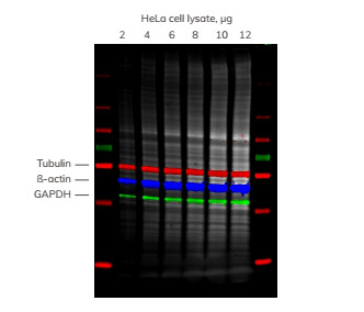
Ponceau S is a fast, reversible protein stain that is commonly used to confirm whether a protein sample has been successfully transferred from the gel to the membrane, and the researchers then perform an immunoblotting process. Staining the film after transfer can quickly and easily identify some transfer problems prior to incubation of the blot with the antibody, such as incomplete strips, uneven transfer and artifacts due to the presence of bubbles. In addition, the total protein on the stained membrane provides a standard for the standardization of total protein. Figure 4. AzureRed images simultaneously with three proteins of interest. Diluted HeLa cell lysate was added to the gel. After transfer, the blots were stained with AzureRed and then the tubulin, ß-actin and GAPDH probes were added without decolorization. Scanning was performed using a Sapphire dual-mode multispectral laser imaging system. Four channels are superimposed and total protein (AzureRed staining) is shown in grey; tubulin-red, ß-actin-blue and GAPDH-green Biologics are drugs made from complex molecules manufactured using living microorganisms, plants, or animal cells.Anti-rabies Vacine for Human is a preparation of rabies fixed virus CTN-1V inoculated into Vero cell , which is used for the prevention of the rabies disease. Biological Product,Rabies Symptoms Human,Human Rabies Symptoms,Human Rabies Signs Changchun Zhuoyi Biological Co., Ltd , https://www.zhuoyibiological.com
However, although Ponceau staining is reversible, it is incompatible with fluorescent Western blot detection. Even after thorough decolorization, Ponceau red staining leaves autofluorescence residues on the membrane, increasing background fluorescence.
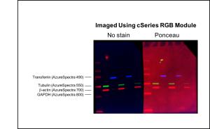
The figure below shows a multicolor fluorescent protein blot. The right half was stained with Ponceau and then decolorized prior to immunoblotting. 
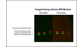
The figure below shows the same footprint taken with the Azure cSeries NIR module, which evaluates the 700nm and 800nm ​​channels. The fluorescent background was reduced in the NIR imaging blot, but still had a high background compared to the unstained half blot. 
To avoid high background due to Ponceau staining, other total protein stains may be considered. 2 AzureRed total fluorescent protein staining is fully compatible with downstream Western blot detection, including fluorescence detection and downstream mass spectrometry.
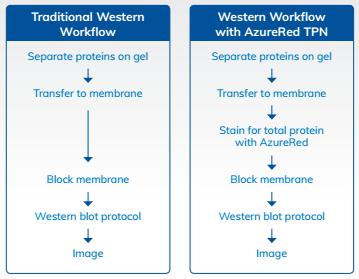
AzureRed fluorescent protein staining is ideal for quantitative western blotting. The wide linear dynamic range ensures that AzureRed is an effective standard for addressing protein loading changes. A key feature of AzureRed is the simplicity of incorporating it into existing western blotting workflows (Figure 3). 
AzureRed dyes can be detected simultaneously with commonly used fluorescent probes without interference, allowing multiple proteins to be evaluated on a single blotting membrane. AzureRed allows standardization of total protein for multicolor blots without the need for stripping and discoloration, allowing multiple proteins to be quantified on a single blot (Figure 4). This method is fast and can be imaged together with the protein of interest. For more information on AzureRed fluorescent protein staining, please visit 
The advantage of total protein stain instead of Ponceau S for fluorescent Western Blots
The two half blots were then blocked with Azure Fluorescent Blot Blocking Buffer and four proteins were detected using four different fluorescent probes as indicated in the figure. The blot was imaged using the Azure cSeries RGB module, and despite complete decolorization, a very high fluorescent background was seen on half of the blots stained with Ponceau.
Figure 3. Total protein normalization using azurered is easy to fit into any Western blotting workflow