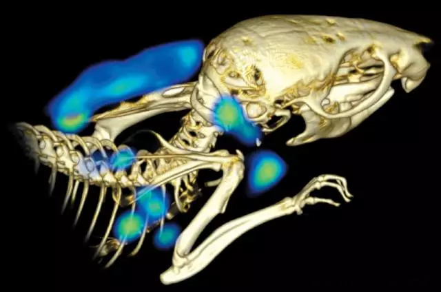
Positron-emission tomography (PET) imaging using antibodies can help researchers observe potential cancer locations in mice and other animals. New technologies help researchers understand the mechanisms of the immune system. The development of anticancer drugs is very tortuous: at first, the prospects of cell experiments and mouse experiments are very optimistic; however, subsequent monkey experiments are very frustrating: monkeys are targeted at targeting and killing pancreatic cancer cells. The drug was poisoned. The drug's R&D team member, Simon Williams of Genentech, Calif., said the team tested the collected tissue samples but found no signs that the drug was toxic. When the researchers imaged the living body and tracked the spread of the drug in the animal, they finally found the crux: the antibody-based drug was mainly absorbed by the bone marrow of the animal, which killed the white blood cells of the bone. In view of this, the researchers gave up the drug. When biopharmaceuticals enter the living, researchers often don't know what will happen next. From early trials to final clinical use, they are not sure how effective the drug is. Sometimes patients respond to drugs; sometimes they don't. Regardless of the outcome, the researchers want to know why. But usually they lack the right research tools. Imaging scientists and cancer researchers are now trying to solve this problem. They combined traditional PET (Positron-emission tomography) technology with antibodies and similar molecules to create a new technology called immunoPET. Researchers say that as cancer treatment becomes more precise and complex, tools for assessing efficacy also need to be continuously improved. Modern biotherapy is only available for some patients, but doctors cannot reliably predict which part of the patient is suitable for this treatment. A biopsy can only tell you what happened to a part of a tumor, and immunoPET can provide a snapshot of all the tumors in your body. Traditional PET uses radiotracers to observe human tissue function, while immunoPET uses antibodies to identify target cells. With the proliferation of cancer immunotherapy and the increasing popularity of therapeutic strategies to mobilize the immune system against tumors, there is a growing interest in this emerging imaging technology in the cancer field. But designing an immunoPET imaging probe is not easy. Factors such as radiotracer selection, antibody design, and imaging kinetics require careful consideration. Fortunately, scientists have made some progress. Now they can identify an increasing number of immune cells and cancer tissues and are adapting antibody structures to improve their properties. New treatment and imaging strategies are on the horizon. Sam Gambhir, director of radiology at Stanford University and a molecular imaging researcher dedicated to early detection and management of cancer, points out that this "immune toolbox" is necessary. Most of the treatment interventions they do are blindly shot, not knowing if the treatment is effective, especially in the early stages. Can people only see if the tumor really shrinks? But if you don't shrink, you don't know what went wrong. This means that people may not know what to do next. Colostomy Bag,Bowel Bag,Colostomy Pouch,Colon Bags Wenzhou Celecare Medical Instruments Co.,Ltd , https://www.celecaremed.com