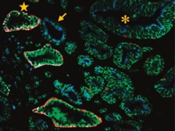
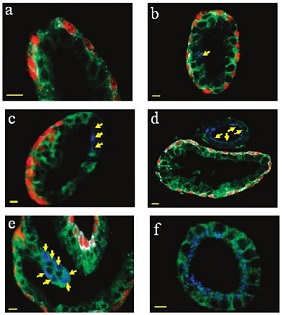
Tumor heterogeneity is a huge challenge in the study of the mechanism of tumor development and the exploration of tumor cell eradication therapy. Tumor heterogeneity is widespread in a variety of tumors (especially breast cancer and prostate cancer are highly heterogeneous), but detection and evaluation methods are still lacking. Current techniques such as RT-PCR, gene chips, protein chips, two-dimensional gel electrophoresis, biomolecular mass spectrometry, etc., result in loss of cellular and histomorphological information. The quantum dot's own optical properties and multi-color imaging capabilities are very suitable for tumor heterogeneity research, and can integrate molecular expression with histomorphological observation, which is of great significance for accurate diagnosis. Professor Shuming Nie of Emory University, for paraffin-embedded tissue specimens of radical prostatectomy and needle biopsy, for four prostate cancer markers E-cadherin, high-molecular-weight cytokeratin (CK HMW), p63, A-methylacyl CoA racemase (AMACR), using quantum dot-labeled secondary antibodies of different species and emission wavelengths, reveals the progression of prostate cancer based on wavelength-resolved fluorescence imaging and spectral analysis by two rounds of primary/secondary sequential staining. Among them, accompanied by changes in glandular structure (transition from basal and luminal cell bilayers to malignant cell monolayers), different molecular marker expression patterns are presented. Compared with traditional tissue section HE staining and immunohistochemistry (IHC) methods, quantum dot multicolor imaging is highly reproducible and has a good correlation with HE and IHC results. It can be expected that for complex and suspected atypical tissue samples, quantum dot multicolor imaging provides integrated information on molecular expression profiles and histomorphology; at the same time, rare cell types (such as cancer stem cells) are detected from complex cell/tissue environments. , a single malignant transformed cell, etc., early detection of the malignant transformation of cells, is of great significance for accurate diagnosis and treatment. Figure 1. Quantum dot multicolor imaging analysis of prostate tumor tissue sections showing tumor heterogeneity. Star-marked normal prostate tissue (expressed in E-cadherin, CK HMW, p63, no AMACR expression), arrow-marked malignant transformed glands (high expression of AMACR, loss of expression of CK HMW and p63), and snowflake labeled prostate intraepithelial neoplasia ( PIN) (the conversion period of both AMACR and p63 expression). Figure 2 Multi-color imaging of quantum dots shows molecular and tissue morphological changes during malignant transformation of the prostate. (a) benign prostate (basal-cavity cell bilayer, AMACR-); (b) single malignant transformed cells (AMACR+) in the luminal cell layer; (c) malignant fragments (yellow arrow, AMACR+) and benign fragments in the prostate (red p63+) and transitional structure (low AMACR expression and p63 deletion); (d) malignant glands 5-10 μm beside the benign gland (yellow arrow); (e) Partial malignant transformation of the pleated glands (yellow arrow); (f) Complete malignant transformation of the gland (high expression of AMACR, loss of expression of CK HMW and p63). Source of the document: Liu J, Lau SK, Varma VA, Moffitt RA, Caldwell M, Liu T, Young AN, Petros JA, Osunkoya AO, Krogstad T, Leyland-Jones B, Wang MD, Nie S. Molecular mapping of tumor heterogeneity on clinical tissue specimens With multiplexed quantum dots. ACS Nano. 2010, 4(5): 2755-65. Tetanus Vaccine,Hepatitis B Vaccine For Adults,Tetanus Booster,Td Vaccine FOSHAN PHARMA CO., LTD. , https://www.fospharma.com
