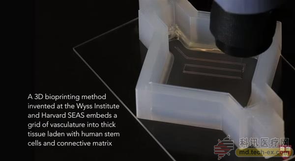
Release date: 2016-03-10 Recently, a team of scientists from Harvard University's John A. Paulson School of Engineering and Applied Science (SEAS) and Harvard's Wyss Bioengineering Institute has invented a method that can use human stem cells, extracellular matrices, and vascular endothelial cells. The circulation channel 3D prints a thick vascularized tissue structure. The resulting vascular network contained within the deep tissue is capable of uniformly infusing liquids, nutrients, and cell growth factors throughout the tissue. This major breakthrough was published in the Proceedings of the National Academy of Sciences on March 7, 2016 . Source: Harvard University End milling cutter For Manual Machine aftermarket
This item showed that our key milling cutter which can use for manual vertical key making machines
Product Features
1.Material: Top quality, high hardness and high wear-resistant carbide, ultra-fine particles, good wear resistance;
2. Manufacturering process: 5-axis automatic grinding center for mirror grinding, sharp edge, low noise; side fixed plane adopts full grinding process, compact installation.
3.Quality: The roundness of the handle is 0.002mm, which greatly reduces the blade yaw during high-speed rotation.
4. Features: accurate size, cutting edge size accuracy of 0.005mm, cutting key size precision; sharp edge
5.Advantages: Specially tested in 994 machine, sharp, can cut brass key and nickel silver key, test Honda original keys, STRATTEC key blanks, smooth edge, no burr, continue to use after processing 200 keys;
6. The Electronic key machine carbide tracer point has good conductivity, ensuring accurate detection, and will not damage the accuracy of the fixture and the spindle.
Key Cutting Blades,car key cutters,key machine cutting wheels ZHANGJIAGANG KEYCUT CO.,LTD , https://www.keycutblade.com
“This latest success extends the ability of our multi-material bioprinting platform to print thicker human tissue, bringing us closer to creating structures that can be used for tissue repair and regeneration.†Senior author of the study, Hansörg Wyss Professor of Bioengineering Jennifer A. Lewis said. 
So far, the biggest obstacle scientists have encountered on the road to building larger human tissues using various cell types is the lack of reliable methods to embed a life-sustaining vascular network into tissues. This is also the significance of the research results of Lewis and his team.
According to Tiangong, based on its previous work, Lewis's team has increased the thickness of 3D printed tissue by nearly 10 times, thus opening up a broad path for the next step of tissue engineering and restoration. This method combines the vascular tube with living cells and extracellular matrices to enable the structure to function like a living tissue. In the study, Lewis and his research team demonstrated that their 3D bioprinting organization can maintain functions like a living tissue structure for more than six weeks!
In the study, Lewis' team demonstrated the process of 3D printing of a one-centimeter thick tissue containing human bone marrow stem cells, which are surrounded by connective tissue. To demonstrate the function of the organization, scientists injected osteogenic growth factors through a supported vascular system and then induced stem cells to develop into bone cells within a month. It is worth mentioning that the vascular system has the same endothelial cells as the real blood vessels. 
"This research will help us build a basic scientific understanding of bioprinted vascularized living tissue," said Zhijian Pei, an official from the National Science Foundation (NSF) who funded the project. "This type of research It will further expand the application of 3D printed human tissue in drug safety, toxicity screening, and ultimately to tissue repair and regeneration."
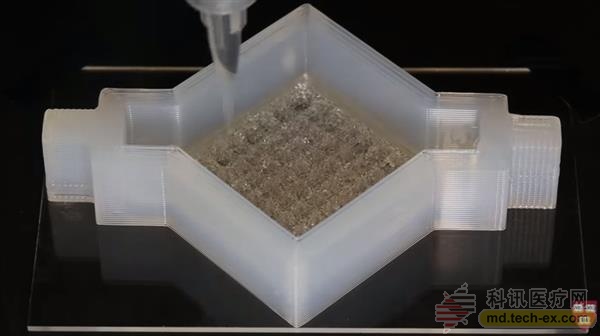
It is understood that Professor Lewis's new 3D bio-printing method mainly uses a customizable 3D printing silicone mold to accommodate and support the printed structure. In this mold, the researchers first print out the vascular line grid and then print the ink containing the living stem cells on it. It should be noted that these inks are self-supporting and strong enough to maintain shape as the size of the structure grows with layer-by-layer deposition. At the intersection of the underlying vascular grid, the researchers print vascular columns that are interconnected to form a ubiquitous microvascular network within the tissue that is deposited throughout the stem cells. After printing, a liquid consisting of fibroblasts and extracellular matrices fills the open area around the 3D printed tissue, cross-linking its entire structure.
The resulting soft tissue is filled with blood vessels, and the researchers can then infuse the tissue with nutrients through the entrance and exit of the silicone mold to ensure cell survival. The ubiquitous vascular system promotes stem cell differentiation by transporting cell growth factors throughout the tissue. For a more intuitive process, please see the demo video below:
Researchers say that if you want to achieve a variety of shapes, thicknesses and compositions of tissue, you can do this by designing the shape of a 3D printed silicone mold and modulating cell inks with different cell types.
"Having this prefabricated blood vessel in the tissue allows us to enhance the deep cell function of the tissue and regulate the function of these cells by injecting nutrients and growth factors, etc." David, one of the first authors of the study Kolesky said.